ABSTRACT
This report describes the author’s experience in an ongoing multi-institutional trial of two alternative therapies for cervical disc disease: conventional anterior cervical discectomy with cortical allograft fusion and plating (ACDF), versus discectomy and replacement with the Prestige™ ST cervical disc device. Because the artificial disc preserves motion at the affected vertebral space, it lessens intradiscal pressures at non-operated adjacent segments. It is hoped that the reduction in stress on adjacent discs will prevent or slow their progressive degeneration.
34 patients were treated at Lancaster General Hospital by the author. All demonstrated clinical and radiographic evidence of single-level cervical disc disease that was causing radiculopathy and/or myelopathy. Patients were evaluated clinically and radiographically before operation, and afterward at 1, 3, 6, 12 and 24 months (36-month follow-up is ongoing). Outcome measures include neurological functional status, visual analog grade of pain, neck disability index, SF-36 general health survey, and radiographic analysis of motion.
After 24 months, all patients showed improvements in all outcome measures, and none had a significantly adverse outcome. Investigational patients who received the Prestige™ ST device all had preservation of motion, and the control patients all had excellent fusion. Although early results showed trends toward improvement in arm pain and neck disability index scores in the investigational group, there were no statistical differences between the groups.
The safety and early clinical results of this study are promising. Longer follow-up is needed to identify important potential differences between these treatment approaches.
INTRODUCTION
Cervical spondylosis is a chronic degenerative condition of the cervical spine that frequently causes debilitating neck and/or arm pain and is a normal part of the aging process.1,2 It affects the vertebral bodies and intervertebral disks of the neck (eg, disk herniation, spur formation), as well as the spinal cord and nerve roots. Symptoms include chronic neck pain, radicular pain, diminished cervical range of motion, headache, myelopathy leading to weakness, and impaired fine motor coordination. The major cause is aging, though injury can also predispose to spondylosis. By age 60 years, 70% of women and 85% of men show radiographic changes consistent with cervical spondylosis.
Conservative management is the mainstay of treatment, and many patients with symptomatic degenerative changes have a satisfactory response to non-surgical interventions. Immobilization of the cervical spine is the primary conservative treatment, and other conservative approaches include cervical traction, targeted exercises, stretching, application of heat, massage, and the use of non-steroidal anti-inflammatory drugs, muscle relaxants, and corticosteroids.
Surgical intervention is usually reserved for symptomatic patients who fail to respond adequately to conservative treatment, and ACDF (anterior cervical discectomy and fusion) has become the surgical treatment of choice 3. The procedure is highly successful in relieving symptoms, has a low rate of associated complications, provides long-term stability, and halts the degenerative process in the treated segment.
One of the major drawbacks of ACDF is the deleterious effect of cervical fusion on adjacent kinematics. Biomechanical studies have shown that cervical fusion increases motion and intradiscal pressures at non-operated adjacent segments, which can increase the stress on adjacent discs and accelerate their degeneration.
Cervical disc arthroplasty by replacement with a prosthetic device has emerged as a relatively new motion-sparing technique for the treatment of degenerative cervical spondylosis.4,5 Unlike ACDF, cervical disc replacement restores normal motion and thus maintains a physiologic biomechanical environment for adjacent disc levels.6 Cervical disc arthroplasty is at least as effective as ACDF in providing symptomatic relief; it also decreases perioperative morbidity, eliminates the need for postoperative external immobilization, and allows earlier return to normal function. Clinical results to date are encouraging, though its long-term efficacy remains unknown.
Several cervical arthroplasty devices are currently available, which vary in materials, range of motion, and techniques for insertion. One of the most well-studied is the Prestige™ Artificial Cervical Disc System (Medtronic Sofamor Danek USA, Inc., Memphis, TN), a stainless steel disc with two articulating components that are attached to the cervical vertebrae with screws. The device provides relatively unconstrained motion, comparable to that of a normal cervical spine segment.
Controlled clinical investigations of the Prestige™ cervical disc began in Europe in 1996, and U.S. clinical evaluations began in 2002 under an FDA Investigational Device Exemption.
This paper briefly describes the development of the Prestige™ cervical disc, and the findings in 34 patients at Lancaster General Hospital who were enrolled in a collaborative nationwide clinical trial that enrolled 500 patients at 25 centers. The LGH cohort was one of the largest groups enrolled by a single surgeon.
EARLY EXPERIENCE
The Prestige™ cervical disc is a refined version of a device developed in the late 1980s by British neurosurgeon Brian Cummins at Frenchay Hospital in Bristol, United Kingdom. 7 The Cummins stainless steel disc featured a ball-and-socket mechanism which was implanted into the intervertebral space and fixed to the anterior cervical spine by bone screws driven into the adjacent vertebrae. Implantation of the Cummins device began in 1991, and most patients experienced favorable outcomes after several years of follow-up.8
The Cummins joint was eventually redesigned to allow more physiologic motion by replacing the hemispherical cup in the lower portion of the device with a shallow ellipsoid saucer. Additional modifications included redesign of the screw-locking mechanism that secured each component to its respective vertebral body and a decrease in the overall bulk of the device.
Prestige Cervical Disc
The next generation of the Cummins cervical disc in 1998 was the Prestige I, ™ which consisted of a lower hemispherical cup that articulated with an upper dome. The major change was the conversion of the socket portion of the articulation to a trough design, which permitted both antero-posterior translation and flexion-extension, much like the normal anatomical condition.
The Prestige I ™ was prospectively evaluated in a pilot study of 17 patients with end-stage disease, most of whom had a history of multiple previous fusion procedures.10 At 24 months, all joints were mobile and antero-posterior translation was preserved. At 48 months there was improvement in all aspects of function and quality of life, with no reported adverse events, and – most importantly - no evidence of symptomatic degeneration of adjacent cervical discs.
The Prestige II™ was developed in 1999 and featured a more anatomic end-plate in which the hemispherical cup of the Prestige I was replaced with an ellipsoidal saucer, and the endplate was roughened to promote bony ingrowth for long-term stability. The design changes allowed more physiologic motion, a reduced profile, and less friction, which minimized the generation of debris.
In “vitro” biomechanical testing was conducted to compare a group of intact human cadaveric cervical spines with another group implanted with the Prestige II™ artificial joint, and a third group treated with simulated ACDF.11 A programmable testing apparatus was used that replicated physiological flexion/extension and lateral bending. The motion of segments with the Prestige II™ disc mirrored the kinematics of the intact spine, whereas the simulated fusion specimens had decreased motion across the fusion site and a potentially harmful increase in motion at adjacent segments. No permanent deformations or failures of the Prestige II™ device were observed during static and fatigue testing9.
Prestige ST™
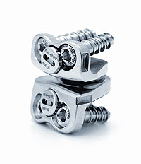
The Prestige ST™ became available in 2002 with a 2-mm reduction in the height of each anterior flange. Its two articulating stainless steel components are attached to the vertebral bodies above and below the disc prosthesis with a constrained locking screw mechanism (Figure 1). Its ball-and-trough design provides relatively unconstrained and physiological segmental motion, and it is available in a variety of sizes. The surfaces that contact the endplates are roughened by grit blasting to promote bony ingrowth. Extensive wear testing of the Prestige ST™ revealed that the total amount of material lost compared favorably with that generated by a cobalt-chromium total hip prosthesis during similar testing.
CLINICAL TRIAL
We recruited 34 patients for a prospective, randomized, controlled study of the Prestige ST ™Artificial Cervical Disc System. The study was conducted at Lancaster Neuroscience and Spine Association, Lancaster, PA, under an Investigational Device Exemption granted by the U.S. Food and Drug Administration.
Inclusion Criteria
The trial was designed to compare the Prestige ST™ Cervical Disc System with a control procedure (standard anterior cervical discectomy with cortical allograft fusion and plating) in the treatment of degenerative cervical disc disease with intractable radiculopathy or myelopathy caused by neuroradiologically documented disc herniation or osteophyte formation . Inclusion criteria consisted of:
- Single-level disease in C4–5 or C6–7,
- Unresponsiveness to nonsurgical treatment or the presence of progressive symptoms or signs of nerve root compression during nonsurgical management,.
- Age greater than 18 years.
We excluded patients with previous surgical treatment of the cervical spine and those presenting with a cervical spine condition other than symptomatic cervical disc disease that required surgical treatment. Patients with osteopenia, osteoporosis, or osteomalacia, were also excluded.
Candidates for the clinical trial received information about the study and were invited to participate. After signing the informed consent document, participating patients were randomized in a 1:1 manner at the study site.
Demographic Variables
Overall, 34 patients were enrolled in the study, 16 in the investigational group and 18 in the control group. There were no significant differences between groups (p > 0.05) in any of the variables assessed, including age, gender distribution, tobacco and alcohol use, race, education level, preoperative use of medications, and duration of symptoms prior to surgery (Table 1).
Preoperative Evaluation
At the time of enrollment, the patients completed several questionnaires: the Neck Disability Index (NDI), the SF-36 general health survey, and a visual analog scale (VAS) for neck and arm pain. In addition, a detailed neurological examination was performed and analgesic requirements, employment status, smoking status, and preexisting medical conditions were determined.
Preoperative radiological studies of the cervical spine included AP, lateral, and flexion/extension studies, in addition to neuroradiological documentation of any compromised nerves. Two independent radiologists reviewed the flexion and extension x-rays of the cervical spine and measured the motion angles at the level to be treated, and at the adjacent levels above and below.
Follow-Up Procedures
Standardized evaluations were performed by the operating surgeon at 1, 3, 6, 12, and 24 months after surgery. Clinical evaluation, with particular attention to neck and upper limb function, included the same radiological studies and measurements of the cervical spine as were done preoperatively. The patients completed the same questionnaires during follow-up as before surgery, and they reported any adverse events.
RESULTS
Radiographic Outcomes
In the investigational group, the mean preoperative angulation at the treated level was 11.09°. This degree of motion was preserved postoperatively with a mean angulation of 10.6° at 12 months, and 9.48° at 24 months. The radiographic outcomes at the treated level were not significantly different in the control group.
The mean preoperative angulations in the segments above and below the treated segment were 11.51° and 10.48° respectively in the investigational group. This motion above and below was also preserved postoperatively, with a mean angulation of 11.23° and 11.41° respectively at 12 months, and 11.93° and 11.58° respectively, at 24 months. The radiographic outcomes at the segments above and below the treated level were similar in the control group. The rate of successful fusion in the control group was 100% at 24 months.
Neck and Arm Pain
At all postoperative intervals, both treatment groups showed significant improvement from preoperative scores (P<.001) for the NDI, as well as neck and arm pain frequency and intensity as assessed by VAS. Statistical equivalence between the treatment groups was demonstrated at each postoperative interval up to the 24-month follow up.
General Health
The investigational group showed improvement similar to the control group in both the physical and mental component categories of the SF-36 at all postoperative intervals. The differences in scores between the treatment groups were not statistically significant.
Rates of Successful Outcomes
Rates of success for gait, arm pain, neck pain, general health, neurologic status, and radiographic outcomes at 24 months are shown in Table 2.
Successful outcomes for gait, arm pain, neck pain, and general health were defined as postoperative maintenance or improvement of preoperative status. Overall neurologic success was defined as maintenance or improvement from preoperative status for motor, sensory, and reflex elements combined. Radiographic success in the investigational group was defined as absence of bridging bone and angular range of motion between 4° and 20°.
Perceptions of Outcomes
The efficacy of treatments was assessed by the patients and the lead investigator at each postoperative interval. At 24 months, 57% of the patients in the investigational group and 33% of those in the control group reported feeling “completely recovered;” 43% vs. 39% respectively reported feeling “much improved;” and 0% vs.28% respectively reported feeling “slightly improved.”
Physician-rated perceptions of outcomes at 24 months in the investigational and control groups were “excellent” in 79% and 39% respectively, “good” in 21% and 52%, and “fair” in 0% and 9%.
Adverse Events
There were no significant differences between the groups in mean operative time, blood loss, duration of hospital stay, or postoperative medication use. No patient in either group required the use of an external orthosis postoperatively. At 24 months there was no evidence of screw migration, fracture, or other type of device malfunction in the investigational group.
DISCUSSION
This study demonstrates that the Prestige ST™ cervical disc provides immediate relief of symptoms comparable to results with ACDF, while maintaining motion at the treated segment without compromising adjacent segments. Most clinical outcome measures trend in favor of the Prestige ST™ disc, although the differences are not statistically significant. The observation period is too short to make any conclusive statements about the benefits of preserving motion in adjacent segments, but the principles of earlier biomechanical studies suggest that it should have a positive influence on the natural history of segment degeneration.
That clinical outcomes between the study groups were comparable in the short term represents a primary success for the new technology, but the real benefits of the device - preservation of motion and elimination of adverse influence on the adjacent segments - are long-term benefits that cannot be assessed before several more years of observation have been completed. ACDF will remain the gold standard of surgical treatment for cervical disc disease until longer term studies confirm that these theoretical advantages of disc replacement translate into enduring clinical benefits.
CONCLUSION
Several cervical disc prostheses are currently in various stages of development and clinical usage, and it is not known which device is ideal. The preliminary clinical experience gained with the Prestige ST™ cervical disc in well-designed prospective studies provides an important foundation for the eventual incorporation of this methodology into clinical practice. Further clinical trials, larger sample sizes, longer follow-up, and greater statistical power are needed to confirm the preliminary results of the present study, and such studies are currently in progress. Reports of additional follow-up in patients who receive the implants are eagerly anticipated.
REFERENCES
- Shedid D, Benzel EC. Cervical spondylosis anatomy: pathophysiology and biomechanism. Neurosurgery 2007;60(1 Suppl 1):S7-S13.
- Orr RD, Zdeblick TA. Cervical spondylitic myelopathy. Approaches to treatment. Clin Orthop Relat Res 1999;359:58-66.
- Pracyk JB, Traynelis VC. Treatment of the painful motion segment. Cervical arthroplasty. Spine 2005;30:S23-S32.
- Acosta FL Jr, Ames CP. Cervical disc arthroplasty: general introduction. Neurosurg Clin N Am 2005;16:603-607.
- Phillips FM, Garfin SR. Cervical disc replacement. Spine 2005;30(17 Suppl):S27-S33.
- Wigfield C, Gill S, Nelson R, et al. Influence of an artificial cervical joint compared with fusion on adjacent-level motion in the treatment of degenerative cervical disc disease. J Neurosurg 2002;96:17-21.
- Traynelis VC. The Prestige cervical disc replacement. The Spine Journal 2004;4:310S-314S.
- Cummins BH, Robertson JT, Gill SS. Surgical experience with an implanted artificial cervical disc. J Neurosurg 1998;88:943-948.
- Wigfield CC, Gill SS, Nelson RJ, et al. The new Frenchay artificial cervical joint. Results from a two-year pilot study. Spine 2002;27:2446-2452.
- Robertson JT, Metcalf NH. Long-term outcome after implantation of the Prestige I disc in an end-stage indication: 4-year results from a pilot study. Neurosurg Focus 2004;17:69-71.
- DiAngelo DJ, Robertson JT, Metcalf NH, et al. Biomechanical testing of an artificial cervical joint and an anterior cervical plate. J Spinal Disord & Techniques 2003;16:314-323.
Perry Argires, M.D., F.A.C.S.
Lancaster Neuroscience & Spine Associates
1671 Crooked Oak Drive
Lancaster, PA 17601
717-569-5331
pargires@lancspine.com
Table 1. Demographic Information
|
Variable
|
Investigational
(N=16)
|
Control
(N=18)
|
p-value *
|
Age (yr.)
Mean ± SD
Range |
43.9 ± 7.2
34.0-58.0 |
43.3 ± 8.8
29.0-60.0 |
0.831 |
Height (in.)
Mean ± SD
Range |
66.9 ± 4.1
59.0-72.0 |
67.1 ± 4.4
58.0-75.0 |
0.874 |
Weight (lbs.)
Mean ± SD
Range |
192.9 ± 41.2
125.0-275.0 |
182.6 ± 37.1
117.0-255.0 |
0.447 |
Sex [n (%) ]
Male
Female |
8( 50.0)
8( 50.0) |
9( 50.0)
9( 50.0) |
1.000 |
Race [n (%) ]
Caucasian
Hispanic |
15( 93.8)
1( 6.3) |
18(100.0)
0( 0.0) |
0.471 |
Marital Status [n (%) ]
Single
Married |
3( 18.8)
13( 81.3) |
2( 11.1)
16( 88.9) |
0.648 |
Education Level [n (%) ]
< High School
High School
> High School |
1( 6.3)
8( 50.0)
7( 43.8) |
1( 5.6)
5( 27.8)
12( 66.7) |
0.487 |
Tobacco Used [n (%) ]
Yes
No |
4( 25.0)
12( 75.0) |
4( 22.2)
14( 77.8) |
1.000 |
Alcohol Used [n (%) ]
Yes
No |
10( 62.5)
6( 37.5) |
10( 55.6)
8( 44.4) |
0.738 |
| *For continuous variables, p-values are from ANOVA and for categorical variables, they are from Fisher's exact test. |
Table 2. Summary of Success Rates (24 Months)
|
Variable
|
Investigational
(N=16)
|
Control
(N=18)
|
Arm Pain [Number (%) of patients]
Success
Failure |
14(100.0)
0( 0.0) |
17( 94.4)
1( 5.6) |
Neck Pain [Number (%) of patients]
Success
Failure |
13( 92.9)
1( 7.1) |
18(100.0)
0( 0.0) |
Gait [Number (%) of patients]
Success
Failure |
14(100.0)
0( 0.0) |
18(100.0)
0( 0.0) |
| Neurologic Status [Number (%) of patients] |
|
Motor
Success
Failure
|
14(100.0)
0( 0.0) |
17( 94.4)
1( 5.6) |
|
Sensory
Success
Failure
|
14(100.0)
0( 0.0) |
18(100.0)
0( 0.0) |
|
Reflexes
Success
Failure
|
14(100.0)
0( 0.0) |
18(100.0)
0( 0.0) |
|
Overall
Success
Failure
|
14(100.0)
0( 0.0) |
17( 94.4)
1( 5.6) |
SF-36 General Health Status [Number (%) of patients]
Success
Failure |
8( 57.1)
6( 42.9) |
11( 61.1)
7( 38.9) |
Radiographic Status [Number (%) of patients]
Success
Failure |
9( 81.8)
2( 18.2) |
N/A
N/A |
Back to Top
Figure 1. Prestige ST™