Fall 2023 - Vol. 18, No. 3
PHOTO QUIZ FROM URGENT CARE
Adolescent with a Rash
Mary Kathryn Flory, FNP-C
Nurse Practitioner
Penn Medicine Lancaster General Health Urgent Care
CASE HISTORY
A 12-year-old and his father present to Urgent Care with a rash that began several weeks prior. Over-the-counter medicated ointments and creams have been applied to the skin with no improvement.
The patient states that the rash is painful and itchy. Initially the rash started on the bilateral wrists, however it is now located on the bilateral lower extremities as well; several areas are weeping. Dad says the rash was initially red with a dry, scaly appearance. The patient has not had a fever and denies recent cold or flu-like symptoms. There have been no known exposures to poison ivy, poison oak, or poison sumac. No one else in the home has a similar rash.
The patient has no known allergies to foods or medications, and his dad denies any new detergents or soaps in the home. The father also denies any history of dermatology problems for the patient.
On physical examination:
- Vital signs are within the normal range and unremarkable.
- The patient is not ill appearing.
- The bilateral wrists have erythematous papules and pustules, some of which are crusted over (see Fig. 1).
- The bilateral lower legs are notable for widespread pustules and patches of brown and honey-crusted lesions with erythematous bases present (see Figs. 2 and 3).

Fig. 1. The bilateral wrists reveal erythematous papules and pustules, some of which are crusted over.
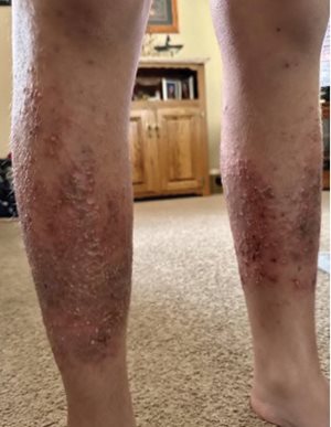
Fig. 2. The anterior view of the bilateral lower legs shows widespread pustules and patches of
brown/honey-crusted lesions with erythematous base present.
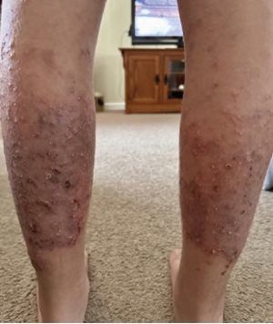
Fig. 3. The posterior view of the bilateral lower legs shows widespread pustules and patches of
brown/honey-crusted lesions with erythematous base present.
QUESTIONS
- What is the differential diagnosis?
- What is the most appropriate diagnosis?
- How should this condition best be treated?
- When is the patient no longer considered contagious?
ANSWERS
- The differential diagnosis might include: eczema with secondary infection; atopic or contact dermatitis; bacterial infection, such as impetigo; viral exanthema from a variety of causative factors, including varicella, herpes simplex, and coronavirus; scabies or lice; candidiasis; or autoimmune disorders such as discoid lupus.
- The most appropriate diagnosis in this case is impetigo, typically caused by staphylococcus or streptococcus species.
- Treatment for localized impetigo could simply entail topical mupirocin 2% ointment or cream applied three times daily for seven days. However, since this patient’s reaction is widespread, oral antibiotics are indicated for treatment; first-line treatment would begin with cephalexin 500 mg four times a day for seven days. Dicloxacillin, doxycycline, or trimethoprim/sulfamethoxazole could be prescribed as well. Erythromycin or clindamycin should not be used because of emerging resistance.1 It is unclear whether over-the-counter agents such as tea tree oil have enough utility to recommend as treatment.
- Typically, the patient is no longer contagious when the individual has been on oral or topical antibiotic treatment for 24-48 hours. In addition, the patient’s lesions should be crusted over and no longer weeping.1
DISCUSSION
Impetigo is a highly contagious superficial skin infection that can occur in any age group but is more common in the preschool population. The findings in patients with non-bullous impetigo include lesions with a flat erythematous base or maculopapular lesions that progress to small pustular lesions. The eruption of the pustules produces the classic honey-crusted plaque (as seen in Fig. 2). Bullous impetigo typically presents as vesicles that form fluid-filled flaccid bullae; when the bullae rupture, a shiny erythematous base with scaling and a brown- or gold-crusted plaque can appear.
2-4
Staphylococcus aureus, Streptococcus pyogenes, or a combination of both causes non-bullous impetigo, commonly called impetigo contagiosa.
Staphylococcus aureus is the pathogen that causes bullous impetigo; this variant is also known as impetigo herpetiformis. First-line treatment is topical: mupirocin or retapamulin. However, if the patient has widespread impetigo, oral antibiotics are warranted and more practical than topical treatment.
1
While topical and oral therapy is important, it is imperative to prevent the spread of impetigo through general hygienic practices. Frequent handwashing, cleaning the lesions with soap and water at least twice a day, bathing or showering regularly, and keeping the sores covered can help decrease the spread of impetigo. The American Academy of Dermatology Association recommends a small amount of bleach (less than a capful) in a bathtub full of an individual’s bath water if recurrent impetigo is evident. Washing bedding and towels separately in hot water and cleaning counters, doorknobs, and other surfaces can also prevent transmission. Avoiding skin-to-skin contact with an individual diagnosed with impetigo is recommended until the patient is no longer contagious, which would typically be when the sores are crusted over or once the patient has been treated with antibiotics for 24-48 hours.
2,5
REFERENCES
1. Hartman-Adams H, Banvard C, Juckett G. Impetigo: diagnosis and treatment.
Am Fam Physician. 2014;90(4):229-235.
2. Johnson M. Impetigo.
Adv Emerg Nurs J. 2020;42(4):262-269.
3. Stevens DL, Bisno AL, Chambers HF, et al. Practice guidelines for the diagnosis and management of skin and soft tissue infections: 2014 update by the Infectious Diseases Society of America.
Clin Infect Dis. 2014;59(2):147-159.
4. VanRavenstein K, Durham C, Williams T, et al. Diagnosis and management of impetigo.
Nurse Pract. 2017;42(3):40-44.
5. Impetigo: Diagnosis and Treatment. American Academy of Dermatology Association. March 11, 2021. Accessed May 2, 2023.
https://www.aad.org/public/diseases/a-z/impetigo-treatment