Fall 2021 - Vol. 16, No. 3


Lang Mantush
PHOTO QUIZ FROM URGENT CARE
A Cystic Lesion on the Elbow
David Lang, PA-C, MPAs
Kayla N. Mantush, PA-C, MHS
Penn Medicine Lancaster General Health Urgent Care
A 62-year-old male presented to Urgent Care with what he described as a “boil” on his left elbow. He said the lesion started out the “size of a pea” and had been growing progressively for the past year. The lesion was tender and disrupted sleep. The patient attributed it to an injury to the skin 5 years earlier. The lesion had been drained about one year ago in an attempt to remove it, but it had since recurred. There was no other signifcant or relevant medical history.
On physical examination vital signs were normal. There was a 3x3cm mobile, cyst-like lesion overlying the left lateral epicondyle (Figs. 1, 2, 3). There was no drainage, erythema, rash, red streaking, neurological deficit, decreased range of motion, or pain with movement. The lesion was minimally tender to palpation.
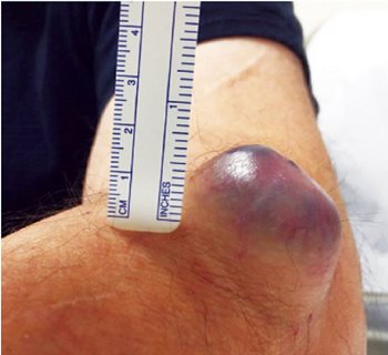
Fig. 1. A 3x3 cm cyst-like lesion was overlying the left lateral
epicondyle.

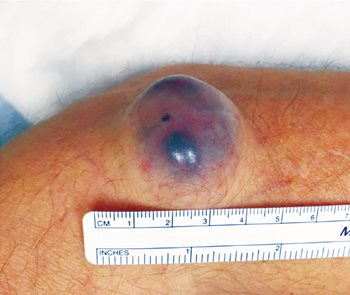
Figs. 2 & 3. The lesion had been growing progressively
from the size of a pea over the past year.
X-rays of the arm revealed no radiopaque abnormalities.
The provider utilized a telederm consultation, and within 24 hours, Dermatology had recommended a biopsy and pathologic examination. The patient was fast-tracked to Dermatology, and they ultimately removed the lesion.
What is the Differential Diagnosis?
Both benign and malignant lesions could be considered, notably:
1. Epidermal inclusion cyst
2. Sebaceous cyst
3. Parasitic cyst (cysticercosis)
4. Pilar (trichilemmal) cyst
5. Hidradenoma
The lesion’s size, location, cystic appearance, and sharply demarcated margins suggest a benign etiology. Epidermal inclusion cysts, sebaceous cysts, and pilar cysts occur most commonly on the head, neck or trunk, not the extremities. Cysticercosis would ordinarily not produce a cyst of this nature and size, and would likely be accompanied by other symptoms.
The consultant from Dermatology made the diagnosis of Hidradenoma.
1
What is a Hidradenoma?
A hidradenoma is a benign adnexal tumor that occurs in both eccrine and apocrine sweat glands with a slightly higher incidence in women. Since sweat glands are ubiquitous, it can present almost anywhere on the body, but most often occurs on the scalp, neck, or limbs. It can present as a solitary, firm nodule or a cyst that typically grows slowly over months or years. Malignant transformation is rare, but can occur. Rarely, lymph node involvement may occur without disease progression.
Histopathology
Hidradenomas can be divided into two patterns – solid or solid-cystic.
2 The most common pattern is the former. The lesions are located in the dermis and can extend into the subcutaneous tissues as encapsulated or un-encapsulated lesions. The cyst wall is most often made up of tumor cells with the cystic cavity possibly caused by the necrosis of tumor cells. The tumor lobules are often covered with collagen fibers, blood vessels, and other interstitium.
REFERENCES
1. Requena L and Sangüeza O. (2017) Apocrine Hidradenoma. In: Cutaneous Adnexal Neoplasms. Springer, Cham.
https://doi.org/10.1007/978-3-319-45704-8_10
2. Li ZY and Chang JM. Solid-Cystic Hidradenoma,
Int J Derm and Vener. 2019; 2(3): 191-192. doi: 10.1097/JD9.0000000000000012
https://journals.lww.com/ijdv/fulltext/2019/09000/solid_cystic_hidradenoma.16.aspx