
Spring 2021 - Vol. 16, No. 1
PHOTO QUIZ FROM URGENT CARE
A Woman with Painful Finger Lesions
Emily Maul, PA-C
Penn Medicine Lancaster General Health Urgent Care
CASE HISTORY
A 33-year-old female presented to Urgent Care complaining of painful small blisters at the base of her left index finger. The lesions appeared about 2-3 days ago and have been worsening since. She reports similar episodes in the past, and attributes the previous episodes to spider bites. She denies fevers, but has had chills.
Examination shows vesicular gray lesions on the palmar aspect of the proximal phalanx of the left index finger, with associated tenderness to palpation. There is swelling of the finger, and range of motion is decreased because of pain. There is no fluctuance or crepitus.
QUESTIONS
1) Are the lesions the result of a process that is infectious, inflammatory, circulatory, allergic, or autoimmune?
2) What is the causative agent?
3) What is the treatment for herpetic whitlow?
4) What can cause recurrent outbreaks of herpetic whitlow?
5) If this was seen at the distal finger, what other disease processes should be considered?
ANSWERS
1) The characteristic appearance, location, and recurrent nature of the lesions, and the lack of external trauma or circulatory compromise, make the likely diagnosis a herpetic whitlow.
1
2) Herpetic whitlow is not a bacterial infection; it is caused by Herpes simplex virus types 1 and 2.
2
3) For the first outbreak, conservative treatment is appropriate, using anti-inflammatories, rest, and elevation. For recurrent infections in immunocompetent patients, oral acyclovir can be used.
1
4) Recurrence of herpetic whitlow can be caused by various stressors including illness, fever, sun exposure, psychological stressors, or menstruation. Typically, recurrent infections are milder, and have a shorter duration.
1
5) At the distal finger, herpetic whitlow can be confused with a felon or paronychia.
2
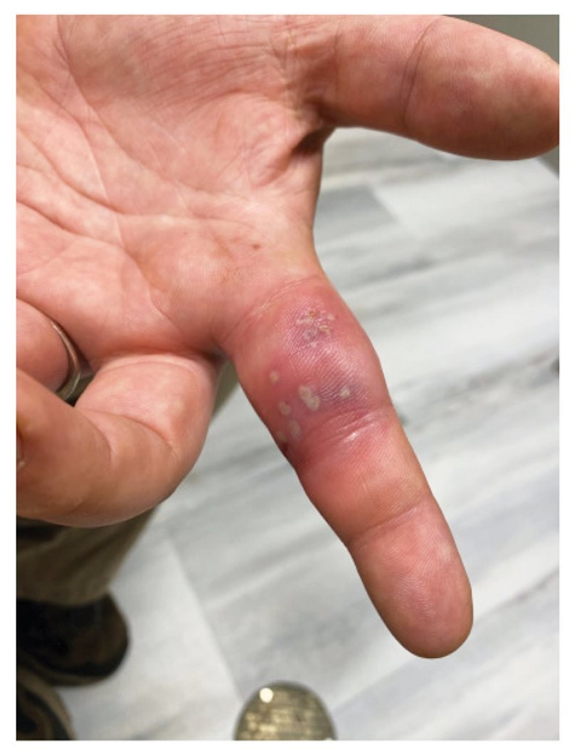
Fig. 1. Patient with herpetic whitlow affecting the proximal phalanx of the
left index finger.
DISCUSSION
Herpetic whitlow is characterized by the formation of multiple erythematous and painful vesicles on a finger (Fig. 1). Usually only a single finger is affected. The vesicles initially are clear, but may appear purulent as the disease progresses. If the vesicle is unroofed, non-purulent fluid may drain. The underlying tissue has a “honeycomb” appearance which is diagnostic for herpetic whitlow. The diagnosis can usually be made clinically, but a Tzanck smear or viral culture can be used to confirm the diagnosis.
1
In children, it typically occurs after the finger comes in contact with the mouth, where it is exposed to HSV due to herpes stomatitis or labialis.
3 It is typical for pain to be out of proportion to the appearance on examination. Patients may have systemic flu-like symptoms.
1 If left untreated, it will typically resolve within 3 weeks.
It is common for this lesion to be misdiagnosed as a bacterial infection, but antibiotics are typically not necessary unless secondary bacterial infection is suspected.
1
REFERENCES
1. Muttath SS, Chung KC, Ono S. Overview of hand infections. UpToDate. Literature review current through January 2021.
https://www.uptodate.com/contents/overview-of-hand-infections
2. Rubright J, Shafritz A. The Herpetic Whitlow.
J Hand Surg. 2011; 36(2):340-2.
3. Patel R, Kumar H, More B, et al. Paediatric recurrent herpetic whitlow.
Case Reports. 2013;2013:bcr2013010207.