Spring 2020 - Vol. 15, No. 1

PHOTO QUIZ FROM URGENT CARE
Traumatic Foot Blisters
Tyler R. Mellinger, MS, PA-C
Penn Medicine Lancaster General Health Urgent Care
CASE HISTORY
A 64-year-old male walked into the urgent care facility for evaluation of pain in his left ankle and foot. The day before he had slipped while getting into a kayak, and immediately developed pain. Nonetheless, he continued to kayak and subsequently was able to ambulate.
The next morning, before presenting to our facility, he had noticed two blisters on his ankle and one on his foot. The pain had intensified, and his foot had become swollen and stiff, with visible bruising. He denied associated fever, drainage from the blisters, or loss of sensation.
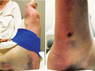
Fig. 1 (left). Medial aspect of left ankle. Two bullae over posterior medial aspect of ankle along Achilles tendon.
Fig. 2 (right). Lateral aspect of left foot. One bulla over proximal fifth metatarsal region.
On examination, he appeared very uncomfortable. Examination of the left lower leg was notable for two bullae over the posterior ankle (Fig. 1), and one over the lateral foot at the base of the fifth metatarsal (Fig. 2). Diffuse edema and ecchymosis were present over the foot and ankle. There was tenderness along the Achilles tendon, and the lateral, proximal foot. The ankle’s range of motion was limited, but he could wiggle his toes and distal sensation was intact and symmetric. Capillary refill was normal. Radiographs of the left foot and ankle were ordered.
QUESTIONS
1. The patient walked into urgent care, should he walk to the X-ray room? What type(s) of injury are you concerned about?
2. What mechanisms of injury increase the likelihood of blister formation over a fracture site?
3. At which sites do these most commonly occur and why?
4. How many fractures does this patient have? (Figs. 3 and 4)
5. What are the next steps in management of this patient?
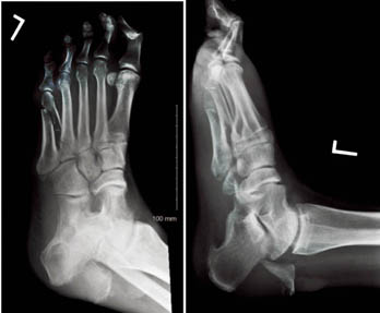
Fig. 3 (left). Oblique X-ray of left foot.
Fig. 4 (right). Lateral X-ray of left foot.
ANSWERS
1. He should not be weightbearing pending results of radiography, as history and exam are highly suspicious for fracture.
2. Fracture blisters are relatively rare, occuring in about 3% of all fractures. They are more likely to occur after high impact injuries.
3. They occur most commonly at anatomic sites where the skin is thin and lacks the underlying protection of muscle or adipose tissue, such as the ankle, wrist, elbow, and foot.
4. There are four total fractures: base of the fifth metatarsal, oblique midshaft of the fifth metatarsal, base of the fourth metatarsal, and calcaneal avulsion fracture with 4.5 cm displacement.
5. Patient should be placed in a bulky short leg Robert Jones dressing with a posterior splint in equinus to relax the achilles tendon. He should be instructed in elevation of the limb, and referred immediately to the care of an orthopedist or to an emergency room. This type of calacaneal avulsion fracture has a high risk of skin necrosis. Additionally, the presence of the overlying blisters complicates care and places the patient at an increased risk of infection.
DISCUSSION
Fracture blisters are an uncommon complication of acute fractures that arise in markedly swollen skin over a fracture. They are tense subepidermal vesicles or bullae filled with fluid or blood. The underlying causative mechanism is a shear injury at the dermo-epidermal junction that occurs at the time of trauma,1 or edema fluid separating the avascular epidermis from the underlying vascular dermis. Proposed contributing mechanisms include: increased interstitial pressure of posttraumatic edema, venous stasis of injured vessles, and localized tissue hypoxia leading to epidermal necrosis.2,3 They are more likely when treatment has been delayed and the extremity has been kept in a dependent position. However, they may also appear after the fracture has been treated, in which case they may only be seen when the cast is removed.
As noted, they are more likely after high energy injuries such as falls from a height and motor vehicle accidents,2 and are more common where the skin is thin without the underlying protection of muscle or adipose tissue. Other risk factors include underlying diseases that predispose to poor wound healing – e.g. peripheral vascular disease, connective tissue disease, hypertension, smoking, alcoholism, diabetes mellitus, and lymphatic obstruction.
Management varies, with some surgeons prefering to leave blisters intact and await resolution prior to surgical stabilization, while others opt for early surgical correction of the underlying fracture without regard for the blister.1,3 Fracture blisters and the underlying soft tissue swelling often delay definitive fracture repair, and if not managed promptly by stabilization and elevation can increase the risk of skin necrosis with chronic ulcers and infection.
REFERENCES
1. Strauss EJ, Petrucelli G, Bong M, et al. Blisters associated with lower-extremity fracture: results of a prospective treatment protocol. J Orthop Trauma 2006; 20:618.
2. Uebbing CM, Walsh M, Miller JB et al. Fracture blisters. West J Emerg Med. 2011;12 (1): 131-133.
3. Varela C, Vaughan TK, Carr J, et al. Fracture blisters: clinical and pathologic aspects. J Orthop Trauma. 1993;7(5):417–427