 Click to Print Adobe PDF
|
Leigh S. Shuman, M.D.
Staff Radiologist, Lancaster General Hospital
Lancaster Radiology Associates, Ltd.
|
 |
An estimated 700,000 symptomatic vertebral compression fractures due to osteoporosis occur each year in the U.S., resulting in 115,000 hospitalizations. They are three times as frequent in women, cause considerable suffering and disability, and since they primarily affect the elderly, will become much more frequent as the population ages. Since conservative management consisting of rest and opiate analgesics has limited success, and causes considerable physical and functional morbidity in older patients, minimally invasive procedures have great appeal.
Percutaneous vertebroplasty (PV) was developed in France in the mid-1980's. The essence of the procedure is the placement of large-bore needles into the symptomatic vertebral body or bodies by a transpedicular approach, followed by injection of methylmethacrylate (bone cement), which stabilizes the fracture and thus relieves pain. A modification of this procedure called percutaneous kyphoplasty (PK) was described in 1998, in which balloons placed through the needles are inflated to reduce the kyphotic deformity and increase the space in the vertebra before the cement is injected. There has been a great deal published about these two procedures over the past decade debating the relative merits and disadvantages of these two approaches. There are no randomized controlled trials comparing the techniques; indeed there are no large trials comparing either technique to placebo. Much of the debate has had more to do with marketing than science. What is clear is that both techniques relieve pain in more than 90 percent of patients, and have a low complication rate in skilled hands. Comparative trials are underway, but it will be a while until data are available. Meanwhile, both techniques work well.
Patients presenting with pain thought to be related to a new compression fracture should receive conservative treatment initially, as some will improve rapidly, but if the patient has a neurological deficit, immediate work-up is essential. Otherwise, if no improvement is seen in the first week or so, consideration should be given to PV or PK. For the initial workup MRI is preferred, as it answers the most important questions by indicating whether there is marrow edema in the compressed vertebra, and whether there is retropulsion (extrusion dorsally) of bone into the spinal canal. In the absence of edema the compression fracture is probably old, and unlikely to benefit from treatment. A large retropulsed fragment is a relative contraindication to either technique due to the risk of pushing the fragment further back during the procedure, compressing the cord or roots. If the patient cannot undergo MRI, the combination of CT and bone scan can also answer these essential questions.
A careful physical exam is vital, to be sure that the site of pain correlates with the compressed vertebra. Back pain is protean, multifactorial, and can occur anywhere in the back. We see many patients whose compression fracture is at L1, but whose pain is all at L5; such a patient is unlikely to benefit from the procedure. Other contraindications include cord compression, coagulopathies, and known allergies to the bone cement.
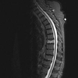 |
| Sagittal inversion recovery MRI sequence of the thoracic spine shows bright white marrow edema in the T7 vertebral body despite minimal loss of height. |
 |
| Fluoroscopic images in a different patient show placement of large bore needles in the L5 vertebral body for cement injection. Patient had previous kyphoplasty at L3 and L4. |
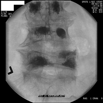 |
| Final distribution of cement in L5 after vertebroplasty. Complete fi lling is not necessary to achieve complete relief of symptoms. |
PV is usually performed on an outpatient basis with conscious sedation. PK is most commonly done in the OR under general anesthesia. It is essential that the placement of the needles and the injection of cement is done under fluoroscopic control, as most of the complications are related either to inadvertent transgression of the canal during needle placement, or spread of cement outside the vertebral body into the epidural space or into veins. Once in the veins, cement can migrate to the lungs. With careful technique, both of these types of complications should be rare.
The procedure typically lasts less than an hour, with the patient kept at rest for several hours post-procedure to allow the cement to harden. Most patients have nearly immediate relief of pain, although some may take several days to realize improvement. A significant risk is the rapid development of new compression fractures at levels above or below the treated sites, as the patient feels much better and resumes more vigorous activity.
Elderly patients with symptomatic compression fractures have endured great suffering over the years, and we will all see more and more patients with this condition, despite efforts to reduce the incidence of osteoporosis. Many patients have benefited significantly from these minimally invasive techniques to relieve their pain.
Leigh S. Shuman, M.D.
Staff Radiologist, Lancaster General Hospital
Lancaster Radiology Associates, Ltd.
P.O. Box 3555
Lancaster, PA 17604
717-544-4900
LSShuman@LGHealth.org